

The Imagined & the Evolved
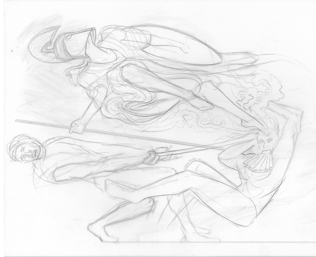
In Hesiod’s Theogony, Zeus experiences extreme symptoms of headache following his deceptive consumption of Metis (goddess of wisdom and insight) whom in a game of transformations he had swallowed—after tricking her to turn herself into a drop of water.
Hephaistos (god of master craftsmanship) attempts to alleviate his father’s pain by cleaving open the head of Zeus with an axe… In this instant, a figure suddenly springs from the head of Zeus bearing a shield and spear.
This was the birth of Athena - Goddess of law, the arts, wisdom and strategic warfare.
The skills for which she is responsible (arguably) number among the most core and (at least in part) most laudable of human pursuits—yet which have nevertheless been the source of innumerate ‘headaches’ across human history…
Anatomical science is the foundation of medicine because it invites us to scrutinise the underlying unity of structures and functions for ourselves. Neuroanatomical observations invite us all to be scientifically informed of how neurobiological structures inform experience: whether these structures are in the everyday flow of constitutional functions which maintain our health, or in clinical circumstances, wherein molecules, genes, cells, tissues or organs may alter in such a way that the adaptivity of these structures is compromised—giving rise to both the symptomatic experience of illness, and signs of disease.
Anatomy and pathophysiology in this way share the common purpose of all sciences—they seek to reveal the underlying reality implied by an observable phenomenon. Migraines are a common yet complex neuropathology, involving an intricate network of neural tissues, and all of which bear a significant responsibility for change among human lives—changes which are at once both proximate (medical) and ultimate (evolutionary).
Craniofacial sensations evolved through deep time to favour the refinement of light touch, caress, thermoception and nociception, alongside other neural modalities that confer significant adaptivity, and to many vertebrate species in continuity with our own, the trigeminal nerve numbers among the most ancient and conserved of all cranial nerves. Early instantiations of the trigeminal nerve itself well precede the evolution of mammaliaformes, and precede human life by hundreds of millions of years - back to the earliest mandibular vertebrates, the gnathosomes 1 from the root: ‘gnathos’ (jaw) + ‘stoma’ (mouth). Hence the trigeminal nerve innervates tissues and reflexes which develop from the 1st pharyngeal arch during embryogenesis—an observable fact of clinical examination today (for example: the masseteric, aka ‘jaw-jerk’, reflex).
In spinal cord tracts and neuropathy, a prior LV article discussed pain as a reflexive and adaptive sensation, illustrating how the gift of sensation in health can be made vulnerable - yielding disease in a system which had otherwise evolved to inform the human experience, and to benefit mammalian life. The underlying origin of the trigeminal nerve within the continuum of animal life, with its comparative anatomy and contemporary medical research, are discussed here in unison—each being essential and intriguing to the context of clinical migraines, and what it means to be human.
Most of all, an understanding of the trigeminal nerve and its functional relationships within the brainstem network can demystify the experience of migraine. Information has its own unique therapeutic value, where the affliction itself might otherwise remain an abstract, unpredictable and frightening source of pain.
Etymology & Structure:
That migraines have afflicted human lives for a considerable period of time, persisting into common parlance throughout cultural history, is denoted by the word migraine itself. Originally from the Ancient Greek noun: hemikrania, which later gave rise to the Latin: hemicranium, and to the English adjective: hemicranial, these terms are commonly used in present day clinical settings to indicate ‘one side / one half of the cranium’.
Linguistically intuitive, the word migraine is derived from a ‘vulgarised’2 French pronunciation of hemicrania, which became phoneticised to ‘micraine’3 by progressing through the 13th and 15th centuries to the present day as migraigne → megrim → migraine. Migraines quintessentially present as a unilateral headache, hence this etymology indicates the word’s use as a shorthand descriptor of localised pain, born heuristically from our experience of the headache phase, this has itself always implied the neuroanatomical structures responsible - however unaware the vast majority of afflicted individuals across human history may have been of their own nervous tissues and organs.
Contemporary scientific investigations into migraine pathophysiology have painted an increasingly complex picture, one which arises largely from the brainstem nuclei and meningeal branches of the Vth cranial nerve (the trigeminal nerve). The nerve itself being the largest (by diameter) of all the cranial nerves, it provides afferent (sensory) innervation independently to each side of the face, including the mucous membranes of the nasal and oral cavities, the sinuses, the ocular adnexa (accessory tissues of the eye: - the cornea, lacrimal glands, conjunctiva).
Importantly this same (ophthalmic) division of the Vth issues a branch from within the cranial cavity which projects to the dura mater: a durable tissue layer which overlays the meninges and meningeal (superficial) vasculature of the brain in the neurocranial vault (figures 3 & 4).
Migrainous pain is regulated and processed centrally by several nuclei in the brainstem (see article 2 in this series) yet part of migrainous pathophysiology indicates a loss of concordant signalling between these nuclei and the meningeal branches of the trigeminal nerve which project into the dura.
As mentioned, the pathways involved in migraine neuropathology are ipsilateral (on one side) to these brainstem nuclei, forming part of a fundamental symmetry of neural structures - which confer to us various adaptive functions of both special and general sensation. The minority of migraineurs who have bilateral aura/visual symptoms have shown functional metabolic activity in both hemispheres, concurring with this relationship between the trigeminal pathways and brainstem nuclei.
Phases & diversity of symptoms:
With increased receptivity by clinical science to patient symptomatology, it is now established that attacks are multi-phasic, marked by one symptom or more for each phase.
Symptoms forming part of the premonitory phase can last for a few hours, to a few days; presenting with a diverse range of afflictions such as fatigue, impaired concentration, mental slowness, neck pain, photophobia, food cravings and/or speech dysfunction.
The aura phase (an ‘aura’ is a transient and focal neurological symptom) lasts between 5 to 6 minutes, and can present with scintillations (twinkling, shimmering or sparkling lights in the field of vision), scotoma (a blind or dark spot in the field of vision), scintillating scotoma (twinkling / arcing lights which are peripheral to / expand into dark spots in the field of vision), paraesthesia (abnormal sensations), numbness of the face/upper limb and/or difficulty in verbalising. Aura occurs in around 1/3rd of migraineurs. A transient wave of neuronal depolarisation in the cortex (cortical spreading depression) is thought to be the underlying pathophysiology leading to symptoms; a ‘silent aura’ is also hypothesised among migraineurs who are asymptomatic for these focal neuropathies.
The headache phase is denoted by the greatest severity of symptoms, lasting between 4 to 72h of the frequently unilateral and pulsatile headache synonymous with migraines;- this phase can incapacitate the individual and worsen with activity. This phase of peak severity can also include nausea, vomiting, photophobia and phonophobia, where 2/> of these as additional symptoms/signs used fulfil the diagnostic criteria for migraine.
The postdromal (recovery) phase can present with asthenia (lack of energy/physical weakness) tiredness, somnolence (drowsiness), difficulty in concentration and with other cognitive tasks4.
Epidemiology:
Migraines afflict 14% of the global population, and around 10 million adults in the UK, at an estimated £8.8 billion in economic cost according to the UK Migraine Trust. They are both more prevalent (3/4:1) and incident among women - and of greater duration and severity, and fall under greater influence from hormonal fluctuations.5 Attacks often begin in puberty, frequently occur perimenstrually, and often cease during pregnancy and whilst breastfeeding.
Among migraineurs, the risk of stroke is extended across both sexes and older age groups, but among women risk is increased considerably if they smoke or use oral contraceptives, both of these having a strong additive effect to this risk. Migraine also shares a bidirectional relationship with depression, where depression increases risk of migraine threefold, and migraine increases risk of depression more than fivefold. Depression also increases the likelihood of migraines becoming chronic (≥15 migrainous days per month, with migrainous characteristics ≥8 days per month for >3 months) according to the British National Formulary.
Introduction to the Vth Cranial Nerve:
The Vth cranial nerve (also known as the trigeminal, semilunar or Gasserian nerve) is a mixed cranial nerve, which predominantly provides afferent (sensory) innervation to cutaneous tissues and mucous membranes on each side of the face, and to the dura mater which overlays the meninges and meningeal (superficial) vasculature of the brain in the cranial vault. Afferent and (limited) efferent (motor root of V) nerve signals are processed centrally by nuclei of the brainstem and the somatosensory cortices.
Along with his contribution as a key founder of the scientific method, the earliest distinction of nervous tissues from tendon and blood vessels by Herophilus (300-250 BC) led him also to discern that motor and sensory functions occur in discrete nerves, and in breaking away from the cardiocentric view of the time, he recognised the brain as the centre of intelligence and sensation. Herophilus must have seen the trigeminal nerve during his dissections, but we only know of his work as an anatomist in Alexandria through Galen and Vesalius. Not until the 16th century were the nerve’s three branches described by Fallopio, and then the nerve named ‘trigeminal’ by Willis. In the mid 18th century, Meckel the Elder consolidated the divisions into the three named branches used today, with the dissection of the ganglion itself being described by Hirsch 27 years later.
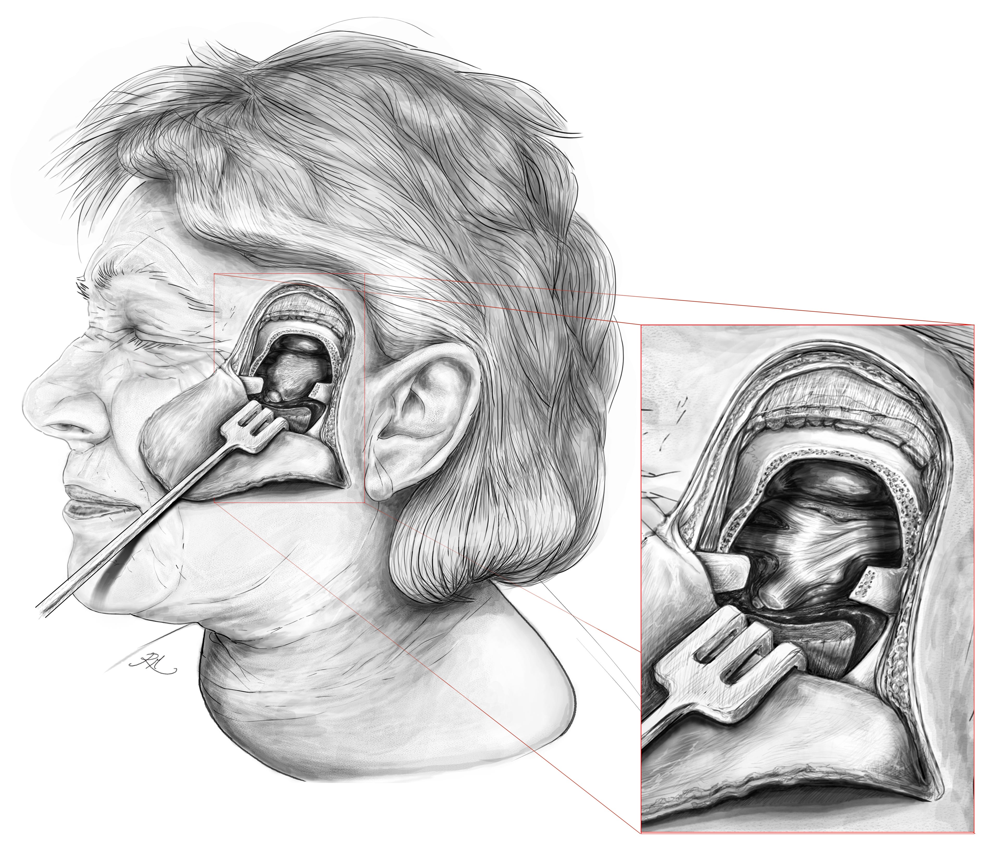
The 1st order neurons which transmit signals from the three sensory regions of the trigeminal nerve have their cell bodies (soma) outside the central nervous system in the trigeminal ganglion.
The ganglion is ensheathed by a glove-like dural pouch (known as Meckel’s cave - see figure 1 above) filled with cerebrospinal fluid. This is continuous with the dura mater of the pons (of the brainstem) through an opening, the porus trigeminus, and like the brain, retains a portion of the fibrous and finely trabeculated membrane (likened to a spiders web) of the arachnoid mater.
The invagination of the trigeminal ganglion by the pia-, arachnoid- and dura-mater (known together as the leptomeninges) offer the same vascular and immunological protection afforded to both the brain and the dorsal root ganglia of the spinal cord, because the thecal layers (meninges) of the spinal cord are continuous with those of the brainstem and cerebrum.
These protective tissues have been conserved by evolution (encoded into the genome of mammalian life) informing what we understand as the everyday functionality of human health. Although confined to the head, V is the largest cranial nerve by diameter, second only to the vagus nerve in extensivity of tissues innervated. The nerve is responsible for thermal, nociceptive, crude, and discriminative sensation in the face, and hence, the neuroanatomy of this nerve can become crucial to medical examination / treatment involving the head and neck.
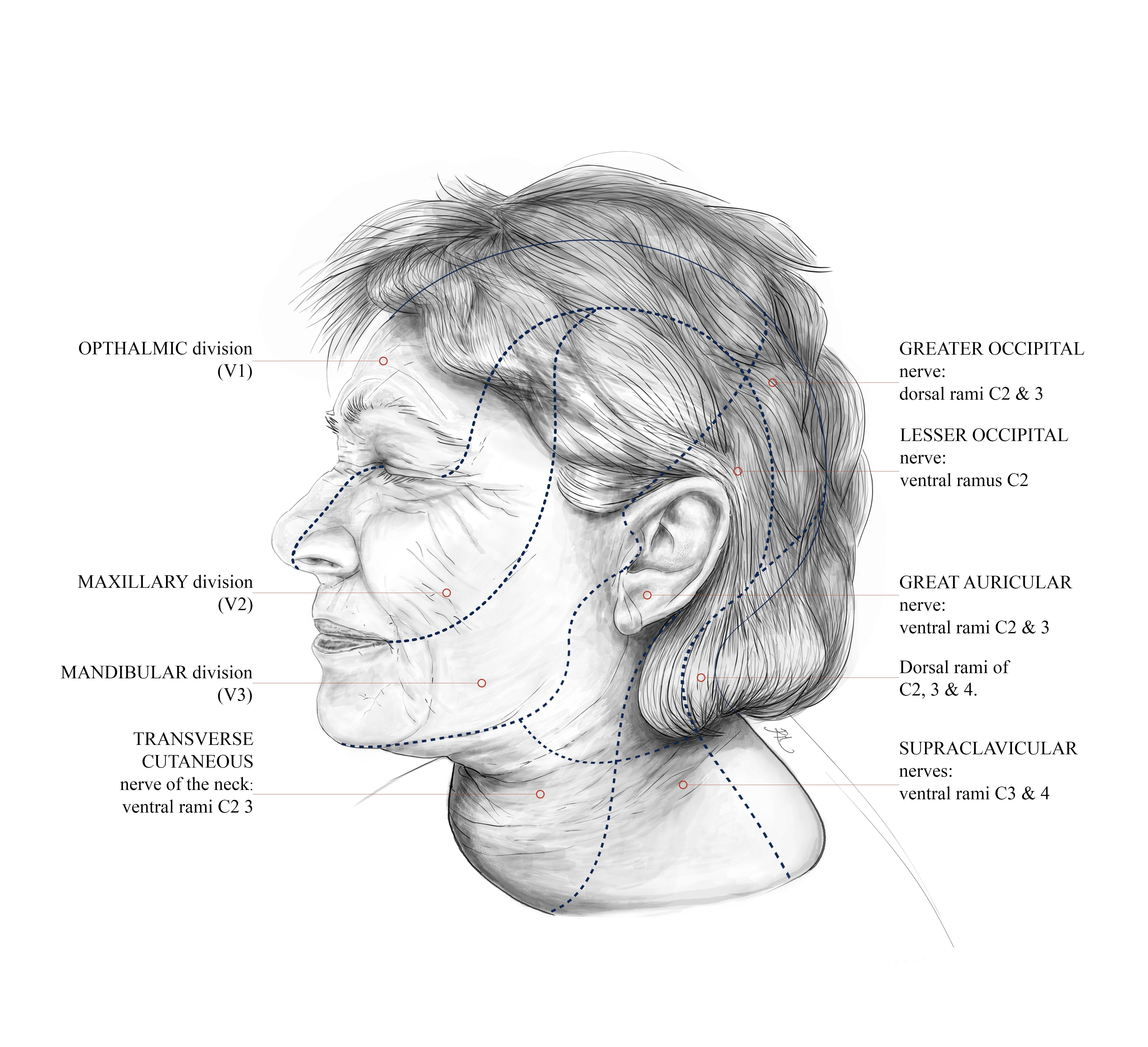
All sensations are initiated within 3 (tri-geminal) segmental zones, demarcated by the three branches of the trigeminal ganglion. When the nerve endings of the skin, accessory nerves to the eye, or mucous membranes are stimulated, their afferent signals transmit to the trigeminal nuclei of the brainstem, only then ascending via 3rd order nerve fibres in the posterior limb of the internal capsule to reach the somatosensory cortices, where these transmissions can be consciously perceived and localised precisely.
The terms division and branch are often used interchangeably, where the former denotes the area of dermatomal (skin) innervation, the latter denotes one of the three nerve branches which issue from the trigeminal nerve.
These branches form nerves, known as: the ophthalmic branch (V1), the maxillary branch (V2), and the mandibular branch (V3).
Sensations from extra-trigeminal areas of the neck and occipital tissues are transmitted to the dorsolateral tract (of Lissauer), and via this tract into the dorsal horn (grey matter) of the spinal cord, where the signal is inhibited if it is below a set neuronal threshold, or otherwise will proceed by the 2nd order and then 3rd order neurons in ascent to the somatosensory cortices of the brain.
Primary sensation transmitted by the nerve from its maxillary and mandibular branches for example, compile sensations transmitted from the oral cavity, allowing for complex textures of food to be differentiated from dynamic motions of fluid.
Humans liquify our food before swallowing it (the reader may have noticed), reducing the mechanical likelihood of the food bolus entering the laryngeal inlet which leads to the lungs (the reader may have also noticed when this does not go as planned).
With a still more expansive view to human development and life history, mastication aids the full gradations and experience of taste, the efficiency of digestion and absorption, and the selection of more complex foods; - factors which have dynamically affected the allometry and morphology of our brains, precipitating human capacities like multi-faceted emotion, imagination, reasoning, logical inference, language and so on.
Bones, Nerves & Symmetry:
For those afflicted with migraines, and by any neuropathology, there is an opportunity for a shared understanding of human adaptation, and where limitations of that adaptation may warrant medical aid.
The question of why nervous tissues in general assume a bilateral symmetry may at first seem rudimentary - if not something we take for granted much of the time in everyday of life.
In considering how crucial this symmetry is (not least for people in need of medical aid for an injury or illness of these tissues) the consideration not only goes to the first principles of how structure and function are developed across the life history of mammals (for in this we are no less dependent on neural structures than they are) but to the quality of life we experience as human beings.
As it remains, the evolution of the trigeminal nerve has been crucial to the human experience of intricate facial sensations. Vital also are the ways in which these sensations complement the special senses (such as sight and sound) and that they inform a plethora of motoric responses by way of peripheral sensation.
Orientation by the head and neck towards stimuli in the outside world for example, not least of which - those solids, liquids and gases which continually make direct contact with the mucous membranes and integumentary tissues of the face.
Sensation is a function of neural structure, through which the trigeminal nerve provides us with our symmetry of perception and response across the integument and mucous membranes of the face: equally in terms of the special senses, having eyes, ears, taste buds and nostrils divided bilaterally (on both sides of the body) maximises sensitivity to the direction and intensity of environmental cues, making our anatomy an adaptive reflection of environmental chemistry and physics. Being no accident that the brain is itself divided into two hemispheres, the spinal cord also assumes its matching symmetry.
Neuropathic pain is typically considered to be a loss of concordant function within specific parts of the central nervous system, and for which the clinical observation of whether the presentation is unilateral or bilateral (by way of symptoms or signs) is medically crucial. In this way, migraine might be considered a loss of such adaptability, and the experience of migrainous pain a physical consequence of this loss.
The trigeminal nerve is estimated to have evolved between 10 to 12 times - among thousands of vertebrate species6.
The 12 cranial nerves give rise to functions of the various sensory viscera, which comprise both the special senses and somatic sensation (external sensation: visual, gustatory, auditory; internal sensation: somatic, enteric, cardiac, proprioceptive etc.).
In order to connect to their brainstem nuclei, all cranial nerves must exit the biochemically and immunologically privileged domain of the brainstem, in which they are intrathecal (ensheathed by the meninges), to subsequently traverse the floor of the cranial cavity through a given foramina, one specially adapted for communication of that nerve from one space to another - between neurocranium and viscerocranium.
Due to these functions being essential for mammalian and human life to survive and thrive, the endochondral conversion of cartilage to bone evolved in order to protect these vulnerable tissues, and to facilitate the foramen and foramina which allow their nerves, blood vessels and muscle to travel from one region to another, including the trigeminal nerve’s connection to its brainstem nuclei centrally, and peripherally to those branches which nociceptively innervate the tissues of the face and dura during migrainous episodes.
Crucial to our understanding of the accommodation of nervous tissues by bone: the term somatic differentiates tissues which are visceral, in terms of gross anatomy. The brain resides in the neurocranium / cranial vault, whilst the cranial nerves and organs reside in the viscerocranium (the ocular cavities, jaw etc). If a nerve is said to have a somatic function, it is responsible for neural innervation of the body and bodily tissues - which have sensory or motoric functions.
The distinction between neurocranial (formed largely via intramembranous ossification: bone converted from fibrous collagen) and viscerocranial tissues (formed largely via endochondral ossification: bone converted from cartilage) of the skull, substantively differentiates the ways in which each of the complex bones of the head form embryogenically, how they function, and how they respond to disease/trauma in life.
The cranial base (‘chondrocranium’) is an exception to how the neurocranial group of bones are generally formed as ‘flat’ (calvarial) bones, being that they result from the endochondral ossification (cartilage to bone conversion), a process which allows for comparably more irregular and fluid structures of bone to boundary, and protect the communication of nerves (+ blood vessels, muscles, membranes etc.) from one anatomical space to another.
Through the most major opening of the neurocranial vault - the foramen magnum, the spinal cord traverses the occipital bone, as well as meningeal branches of cervical nerves (originating from roots C1-C3) that invest into the dura. Conversely, among the most minor foramina of the cranial base, e.g. the foramen spinosum (2-3mm) - the meningeal branch of the mandibular nerve (becoming the nervus spinosus) re-enters the vault, transmitting afferent sensations from the dura mater into the spinal nucleus of the trigeminal nerve (where it resides in the ponto-medullary brainstem).
Largely because the neural integration and processing of our somatosensory functions occur in the brain, the majority of visceral sensations (of the organs) are neurologically referred to the overlying myotome / dermatome; hence ‘referred pain’ is nociceptive sensation issued from an organ - such as the heart, liver or gastrointestinal tract - which can (respectively) be experienced in muscle or skin. This is central to migrainous pathology.
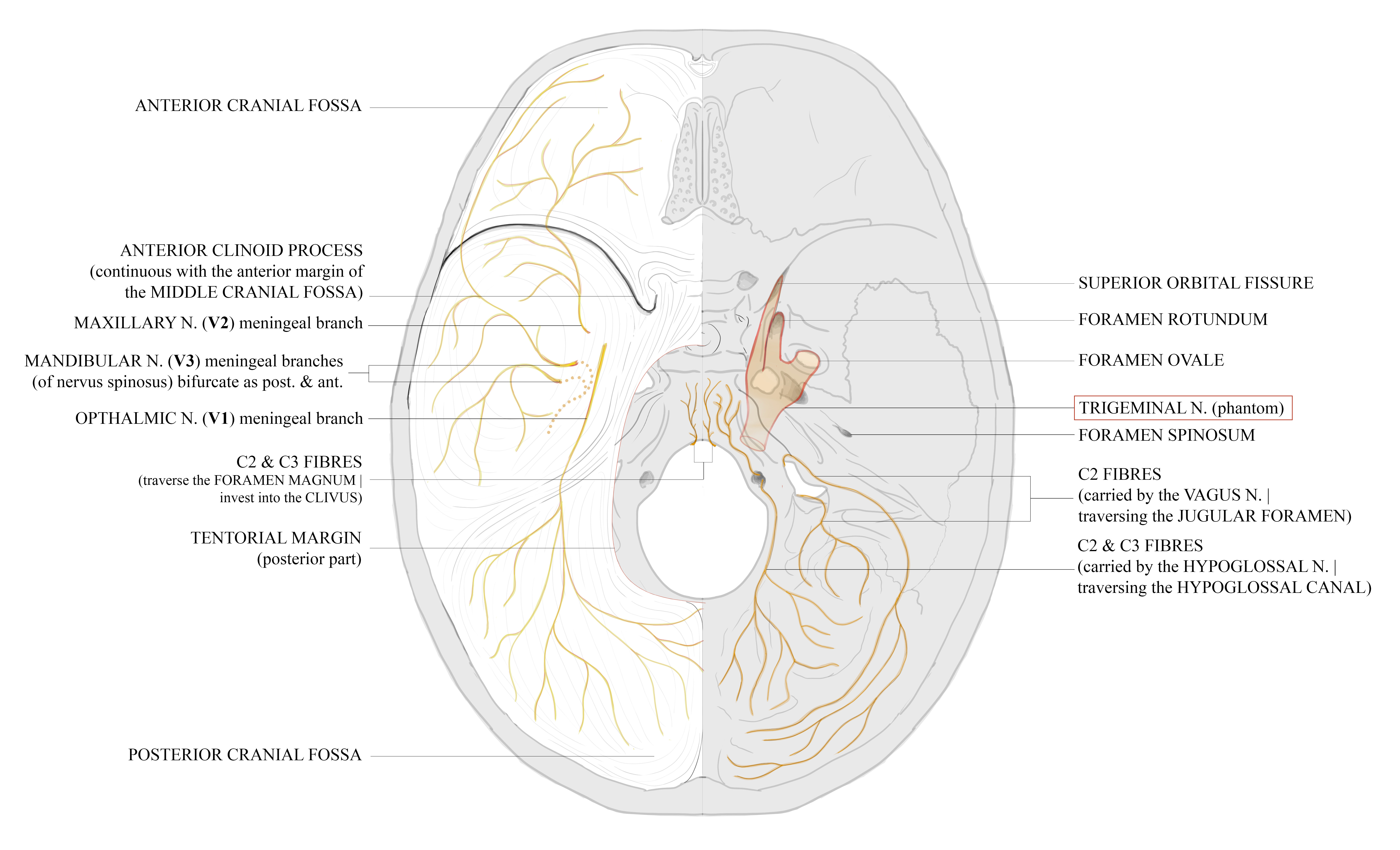
The trigeminal nerve traverses three openings in the middle cranial fossa, as seen in the figure immediately above:
the ophthalmic branch (V1) enters the superior orbital fissure, with its recurrent meningeal branch projecting directly towards the dura mater from within the intracranial space.
the maxillary branch (V2) traverses the foramen rotundum, with its meningeal branch projecting into the dura from within the intracranial space.
the mandibular branch (V3) enters the foramen ovale, and its recurrent meningeal branch re-enters the cranial base via the foramen spinosum.
This meningeal branch of the mandibular nerve is accompanied by the middle meningeal artery (not shown) through the foramen spinosum, with the nerve closely accompanying the proximal trunk of the artery, and as it proceeds, the anterior and posterior divisions of the artery. The nerve fascicles deviate at the periphery however, innervating the dura more broadly as depicted in figure 4.
This neurovascular accompaniment contributes to the neuropathology of migraine because it forms a key part of meningeal innervation by the trigeminovascular network. The trigeminovascular hypothesis has been superseded by the integration of both trigeminocervical + trigeminovascular pathways, giving way to a more integrative view of neuroanatomical function, structure and pathology.
The flat bones of the calvaria (the ‘skull cap’: frontal, temporal, parietal and occipital) develop in accordance with human allometry, in which the neonatal cranium is disproportionate to the rest of the body due to the brain and cranial nerves. Cerebro-visceral development is thus accommodated by the fontanelles of the infant skull, which fuse slowly during infancy to become the cranial sutures - optimising the balance between the intracranial space and the vascular capacity of the central nervous system, which postpartum becomes permanently separated from the maternal blood supply.
The trigeminal nerve resides in the middle cranial fossa, on the anterior surface of the petrous temporal bone. Its divisions are thus accommodated by bone (as shown by the foramen in figure 3) and other tissues, achieving a symmetry optimised for the adaptive sensation of our environment.
Consequential to migrainous pathology, the meningeal branches of V are recursive derivatives of the main ophthalmic, maxillary and mandibular branches; that is, these nerves issue anteriorly from the brainstem, branch into V1, V2 and V3, and then back toward the brain, terminating in the dural layers, where these dura doubly overlay the cranial cavity and the arachnoid mater of the meninges.
Each invested nerve branch is accompanied by an artery, and with this vascular supply the simultaneously durable and periosteal (bone surrounding) dura protects the deeper and more vulnerable layers of the meninges. Only the mandibular branch (V3, see figures 3 & 4) leaves the cranial cavity entirely and then re-enters, effectively communicating twice with the intracranial space.
Less often included in neuroanatomical publications and research (3 sources at time of writing) are all 3 meningeal branches of the trigeminal nerve - which Lucida Visionis has attempted to represent below.
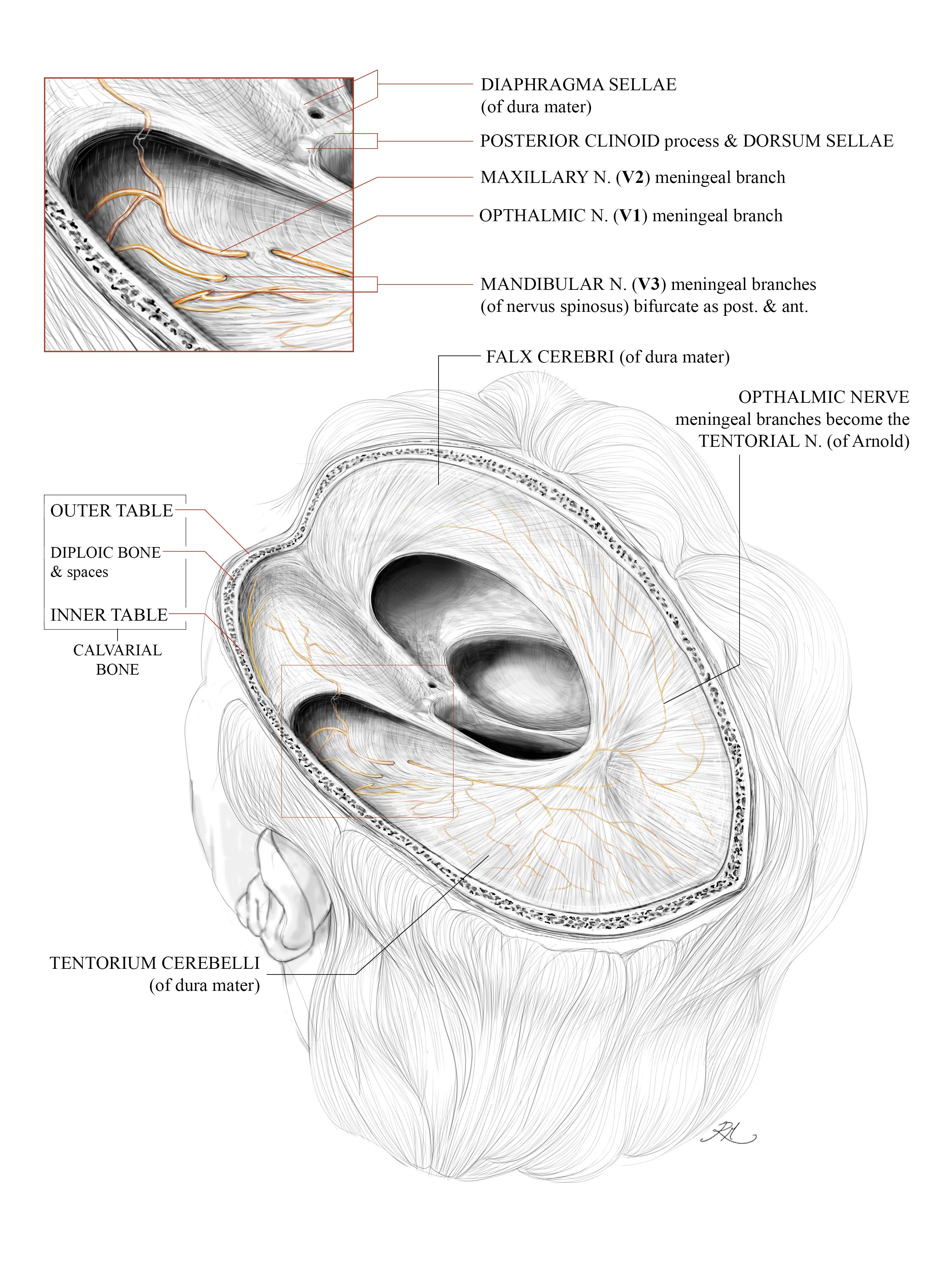
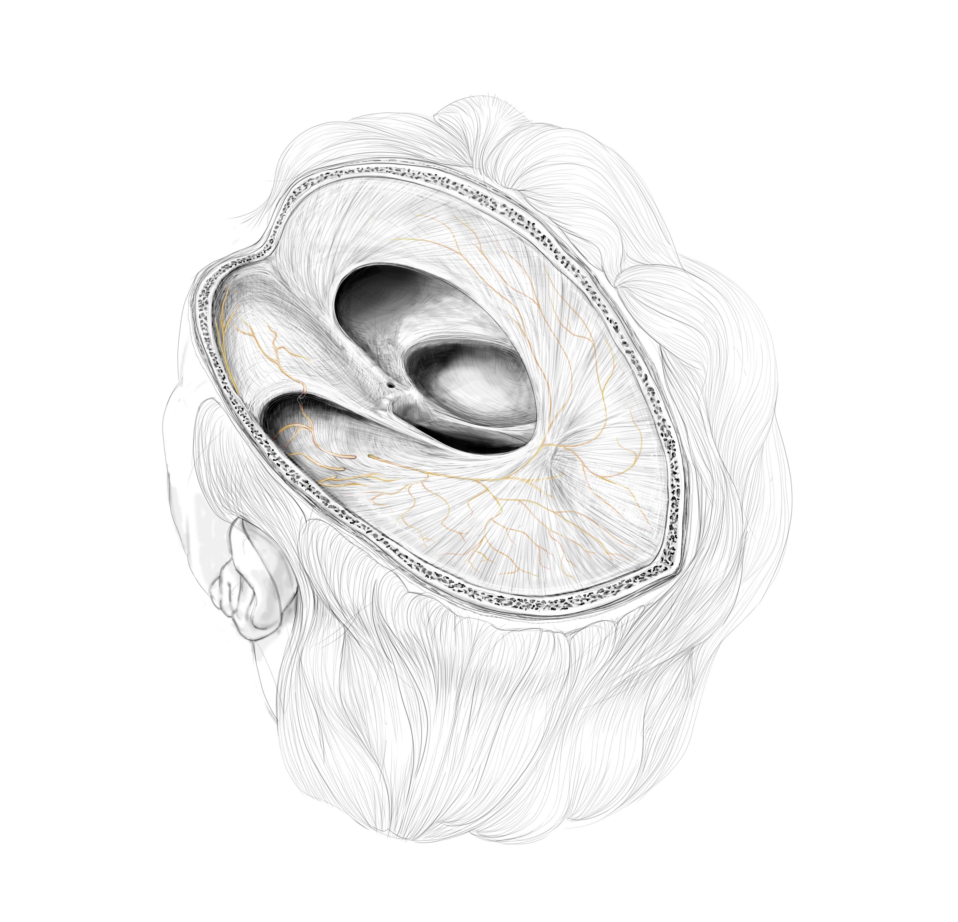
Posteriorly, through the axial plane, meningeal branches from the trigeminal nerve invest into the dura mater (the outermost later of the meninges), emerging across the floor of the cranial vault (as in figure 3) whilst also investing into the dura. The extension of the dura which separates the cerebral hemispheres in its saggital plane is the sickle shaped falx cerebri, and the dura in its axial plane the tentorium cerebelli, which separates the occipital lobes superiorly from the cerebellar lobes inferiorly. The tentorium slopes upwards towards the falx like a marquee, as in the hemicranial dissection shown above.
Margins of the tentorial incisura/notch give way medially to the brainstem, allowing its regions to communicate from the infratentorial (within the tenotorii) space to supratentorial (above the tentorii) space. Along with the entire cranial vault, the incisural space is shown as a true cavity in figure 3 because the cerebra and brainstem have been excised, leaving the u-shaped aperture visible at the centre. The tentorium cerebelli separate the cerebral hemispheres superiorly from the cerebellum inferiorly. The tentorium cerebelli overlay the anterior and middle cranial fossae, and become continuous with the posterior falx cerebri, allowing the communication of the tentorial nerve (of Arnold, as in figure 3) which is the meningeal branch of V1 (the ophthalmic nerve shown in figure 1).
Migrainous craniofacial pain is understood to be an outward physical sign, as nociceptive transmission depends on the meningeal branches of the trigeminal nerve, and their investment into the dura mater of the brain. A greater explanation is required of how the trigeminocervical complex of the brainstem has been shown to inform migraine by processing pain within the trigeminal nuclei, and the regulation of neural pathways which communicate directly and indirectly with the these nuclei of the Vth cranial nerve.
This will be explored in the next article. Thank you for reading Lucida Visionis.
Ashwell KW. Evolution of the trigeminal sensory system and its associated specialisations. In: Kaas JH, editor. Evolution of nervous systems. 2nd ed. Oxford: Elsevier; 2017. p. 433-453. doi: 10.1016/B978-0-12-804042-3.00118-0.
Etymonline. Etymology of migraine [Internet]. [cited 2024 May 22]. Available from: https://www.etymonline.com/word/migraine. You can find the entry at Etymonline.
Oxford English Dictionary [Internet]. Oxford: Oxford University Press; 2025 Dec. migraine (n.), sense 2 [cited 2024 May 22]. Available from: https://doi.org/10.1093/OED/6384918566.
Ferrari MD, Goadsby PJ, Burstein R, Kurth T, Ayata C, Charles A, et al. Migraine. Nature Reviews, Disease Primers. 2022 Jan 13;8(1):2. doi: 10.1038/s41572-021-00328-4.
Sacco S, Ricci S, Degan D, Carolei A. Migraine in women: the role of hormones and their impact on vascular diseases. J Headache Pain. 2012 Apr;13(3):177-89. doi: 10.1007/s10194-012-0424-y.
Tamura M, Ishikawa R, Nakanishi Y, Pascual-Anaya J, Fukui M, Saitou T, et al. Comparative analysis of Hmx expression and the distribution of neuronal somata in the trigeminal ganglion in lamprey and shark: insights into the homology of the trigeminal nerve branches and the evolutionary origin of the vertebrate jaw. Zool Lett. 2023 Dec 5;9(1):23. doi: 10.1186/s40851-023-00222-9.